Derivo Embolization Device for treatment of ruptured intracranial aneurysms
In this month’s issue of The Journal of NeuroInterventional Surgery, an overseas study by , et al evaluates the treatment of 11 ruptured aneurysms with the Derivo Embolization Device (Acandis GmbH & Co. KG, Cologne, Germany) was published. Of the 10 aneurysms available for follow-up, complete occlusion was seen in 90% at 6-months. While this represents a small sample size of patients, it marks another datapoint for the evolving field of flow diversion treatment for cerebral aneurysms. (Derivo not available in the USA.)

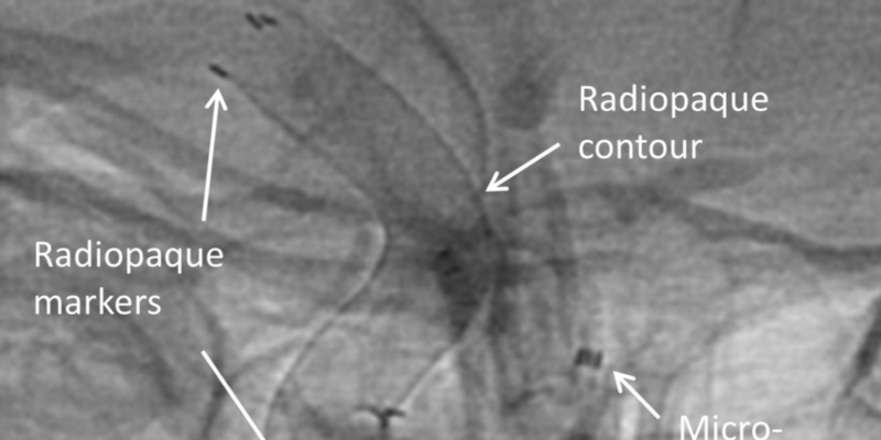
Figure 1 (above) illustrates a middle-aged patient presented with basal subarachnoid hemorrhage (A). Three-dimensional rotational angiography reveals two blister aneurysms (arrows) of the terminal segment of the right internal carotid artery (ICA), represented by two contour irregularities of the vessel wall (B). The blister aneurysms exhibited an unfavorable configuration for coiling and microsurgical clipping. A Derivo Embolization Device (DED) was placed within the ICA covering the affected segment. The unsubtracted images during DED placement show the superior visibility of the device contour and the three radiopaque markers at both ends (C). Digital subtraction angiograms before the procedure (D) and at 6-month follow-up show a complete aneurysm occlusion, whereas the covered side branch remained patent (E).

Figure 2 (above). Native cerebral CT shows basal subarachnoid hemorrhage with blood in the Sylvian fissure and initiating hydrocephalus (arrows) (A). Three-dimensional reconstruction and DSA depict a lobulated superior hypophyseal artery aneurysm of the right internal carotid artery (2.7 mm) (B,C). Owing to irregular aneurysm shape and the extent of subarachnoid hemorrhage, we could not exclude the possibility of aneurysm rupture and a decision for aneurysm treatment was made. Unsubtracted DSA images during Derivo Embolization Device (DED) placement reveal favorable visibility of the device during delivery (D, E). DSA immediately after DED placement shows persistent total aneurysm filling (not shown). At 10-month follow-up, DSA reveals complete occlusion of the aneurysm (F). EVD, external ventricular drain.

Content from this article has been reproduced from “Safety and efficacy of the Derivo Embolization Device for the treatment of ruptured intracranial aneurysms” Goertz L, Dorn F, Kraus B, et al. Volume 11, Issue 3 2019 with permission from J Neurointerv Surg/BMJ Publishing Group Ltd.
Correspondence to this publication’s author can be directed to Dr Lukas Goertz, Center for Neurosurgery, University Hospital of Cologne, Cologne 50937, Germany; [email protected]